Wednesday, August 10, 2011

Malignancy Of Stomach

Patient came with the complain of severe burning  in the epigastric region.
Patient was sent by the physician suspecting  gastritis.
Ultrasonography shows thickened irregular wall of stomach with few small nodes surrounding the stomach.
After water loading, stomach and duodenum  were examined with no outlet obstruction.
There was involvement of gastro-eosophagial junction. 
Gastroscopy and biopsy was done and confirmed the above findings.

Diagnosis - Malignancy of stomach. 
Add caption


Multiple Hydatid Cysts In Liver


Patient came from a village and had a history of handling the animals ( cows & buffaloes) 
She had a lump in right hypochondrium having no complaint of pain
Ultrasonography was done which shows multiple cystic lesions in liver. 
One of the crumpled cyst is with multiple septas.
Big cyst contains few daughter cysts.

Diagnosis :- Multiple hydatid cysts in liver 

 
Add caption


Wednesday, July 20, 2011

Live Tubal Ectopic Pregnancy

Patient presented with acute abdominal pain, mainly on the right side. Clinically it was suspected as acute appendicitis.

Ultrasonography shows normal upper abdomen sonography.
In the pelvis significant free fluid seen with internal echos in the fluid.
Uterus was normal with normal endometrial thickness and very minimal fluid in the endometrial cavity.
Both ovaries were normal. 
In the right adenaxa there was a well defined intact gastational sac with yolk sac and feotal pole in it. 
Real time sonography shows normal cardiac activity in the feotal pole. 
Colour doppler study shows colour flow in the feotal heart.


Add caption


Intra-Occular Dislocation Of Lens

Patient presented with injury in the right eyeball with sudden loss of vision.

Ultrasonography shows variable density echos into the vitreous cavity, suggesting vitreous haemmorrage.


Lens was dislocated and was seen moving in the vitreous cavity.

Add caption


Saturday, July 16, 2011

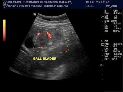
Gall-Bladder Mass

Patient presented with lump in right hypocondrium. 


Ultrasonography shows irregular hypoechoic mass attached to the fundus and body of gall-bladder, not moving in gall-bladder lumen. Calcification seen within the mass. 
Colour doppler study shows significant vascularity, arterial flow in the mass.

Add caption


Arm Lipoma

Patient presented with lump in right arm without pain. 


Ultrasonography shows well defined predominantly echogenic lesion in the subcutaeneous plane with thin strands in it.
Colour doppler study slows no vascularity in the lesion. Underlying muscular plane appear normal.
    Add caption


Breast Cancer

Patient presented with lump in breast. No history of pain. 


Panoramic soft part Sonomammography was done which showed irregular hypoechoic mass with calcification. Colour Doppler study shows increase vascularity in mass. Enlarged nodes were seen in left axilla. 


FNAC was done which confirmed the diagnosis of malignancy of breast.


Add caption